U.S. Department of Energy's Historical Commitment to Improved Healthcare Through Nuclear Medicine
-
1929
Ernest O. Lawrence invents cyclotron
At the University of California's Radiation Laboratory in Berkeley (later to become Lawrence Berkeley National Laboratory), the cyclotron would soon produce the first medically useful radionuclides (iodine-131, thallium-201, technetium-99m, carbon-14, and gallium-67). For this invention, Lawrence will receive the Nobel Prize in Physics in 1939.
-
1946
First delivery of a medical radionuclide to a hospital
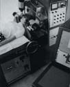
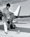
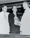 Reactor-produced radionuclides from Oak Ridge become available for medical research. Eugene P. Wigner (in dark suit), director of BER research and development at Oak Ridge, delivers lead-lined container of carbon-14 to Barnard Free Skin and Cancer Hospital in St. Louis. Wigner will receive the Nobel Prize in 1963 for his research on the structure of the atom and its nucleus.
 -
1951
Benedict Cassen invents rectilinear scanner
 Cassen and other BER scientists at UCLA build a scanner that provides images of a thyroid gland based on distribution of an iodine radiotracer, the start of imaging in nuclear medicine.
 -
1952
Hal Anger invents gamma camera
 In Berkeley, California, Anger and his BER colleagues introduce a revolutionary new technique for radionuclide imaging. The gamma camera will become the "workhorse" of nuclear medicine for the next 50 years.
 -
1953
Birth of positron imaging
Gordon Brownell at MIT constructs the first detector device to exploit positron-electron annihilation as an imaging tool, creating a precursor of future PET scanners.
-
1958
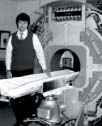
Technetium-99m generator invented
 BER scientists at Brookhaven (Walter Tucker, Powell Richards, and colleagues) invent a generator system that will make Tc-99m the most widely used radionuclide in hospitals worldwide for millions of nuclear medicine patients each year.
 -
1959
Beginning of emission-computed tomography
 David E. Kuhl and other BER scientists at the University of Pennsylvania build the Mark II scanner, ancestor to today's CT and SPECT scanners.
 -
1961
"Headshrinker" direct forerunner of PET
 James S. Robertson, a BER scientist at Brookhaven, develops the "headshrinker," a direct forerunner of PET.
 -
1973
Thallium-201 for medical use
BER scientists at Brookhaven (Elliot Lebowitz, Harold Atkins, and colleagues) develop a faster, more efficient method for producing thallium-201, leading to nuclear stress testing as a routine scan for heart imaging. By the 1990s, doctors will use thallium-201 about a million times a year, accounting for 13% of all nuclear medicine scans.
-
1974
First PET camera built for human studies
 Following several prototypes, Michael E. Phelps, Edward Hoffman, and Michel M. Ter-Pogossian at Washington University, with DOE and NIH support, build the PETT III to use advanced algorithms for computing three-dimensional images.
 -
1976
Development of fluorine-18 FDG for PET
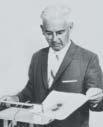
 Alfred P. Wolf (right), Joanna S. Fowler (not shown), Tatsuo Ido (middle), and other BER colleagues at Brookhaven develop and synthesize fluorine-18 fluorodeoxyglucose (FDG), a form of radiolabeled sugar, for PET imaging of glucose metabolism.
First shipment of fluorine-18 FDG to a hospital
 Brookhaven sends F-18 FDG, a PET radiotracer, to the University of Pennsylvania, also a BER research site.
 -
1980
Iodine-131 MIBG for diagnosing and treating rare childhood cancers
New radiopharmaceutical is developed by Donald Wieland and other BER scientists at the University of Michigan.
-
1984
PET image of estrogen receptors in breast tumor
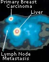 The first PET radiotracer to image a tumor based on a fluorine-18–labeled carrier molecule (fluoroestradiol) that targets a specific hormone receptor of the cell, developed by BER scientists Michael J. Welch (Washington University, St. Louis) and John A. Katzenellenbogen (University of Illinois, Urbana-Champaign).
 -
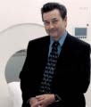
1986
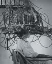
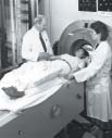 Highest-resolution PET scanner in the world
BER scientists led by Thomas F. Budinger (left) design more advanced PET imaging systems.
-
1987
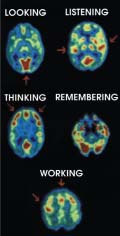 PET
scans show different patterns of glucose (sugar) metabolism related to
performing various mental tasks
PET
scans show different patterns of glucose (sugar) metabolism related to
performing various mental tasksAt UCLA, fluorine-18 FDG PET studies, supported by BER, show different patterns of glucose (sugar) metabolism in the brain during five tasks:
- looking at scenery,
- listening to a mystery story with music,
- thinking by counting backwards from 100 by 7s,
- remembering objects previously memorized, and
- working by touching the thumb consecutively to the four fingers.
-
1998
 Enrico
Fermi Award from DOE
Enrico
Fermi Award from DOEPresidential award is presented to Michael E. Phelps, a BER scientist now at UCLA, for his 1970s work as one of the developers of the first PET camera built for human studies at Washington University, St. Louis.
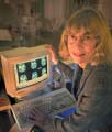 E.O.
Lawrence Award from DOE
E.O.
Lawrence Award from DOEJoanna S. Fowler, a BER scientist at Brookhaven, receives this award for her innovations in development of radiopharmaceuticals and their application for imaging brain chemistry and the biological action of various drugs.
Text adapted from "Converting Energy to Medical Progress" [PDF] (April 2001), Medical Sciences Division, Biological and Environmental Research (BER), Office of Science, U.S. Department of Energy.